Radiology Procedures: Determining Which Scan to Use
When it comes to medical diagnosis and treatment, physicians and other practitioners use a vast range of procedures, methods, and tools in their care.

Radiology Procedures: Determining Which Scan to Use
 15 Dec, 2014

When it comes to medical diagnosis and treatment, physicians and other practitioners use a vast range of procedures, methods, and tools in their care. Radiology plays a major role in the medical arena, and its importance only continues to grow as technology and science contribute continuous new advancements to the field. Radiologists work with a variety of scans, but the most commonly used are CT scan, MRI, general x-ray, and ultrasound. Depending on the nature of the injury or illness, a radiologist may use a single type of scan or use several scans in combination. How does the radiologist determine which scan to use in patient diagnosis and treatment?
A Closer Look at Common Radiology Scans
The x-ray is the most broadly used type of scan, and most people have probably had at least one x-ray in their life; dentists use x-rays to assist them in the proper treatment of patients’ teeth, for example. The x-ray scan uses electromagnetic energy waves to create images of the bones and other interior structures.
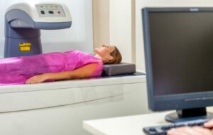
A CT (computerised tomography) scan is a more advanced x-ray which rotates to provide cross-sections of internal images. Using a higher dose of radiation, the CT is able to produce a much more detailed scan, which becomes important when diagnosing or treating certain types of issues.
Ultrasound is well-known for its use in monitoring pregnancy and providing images of the growing foetus inside the mother. Ultrasound technology does not use ionising radiation like the x-ray or CT scan, but uses sound waves to build images of the patient’s body.
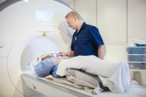
Like the ultrasound, MRI, or Magnetic resonance imaging, does not utilise ionising radiation in its scans. Rather, MRIs work with magnets and radio waves to capture their internal images. An MRI is a scan which takes longer to complete, but the pictures it can produce are highly detailed.
Practical Applications of Radiology Scans
X-rays are very common, particularly for more “visible” ailments such as injuries. For broken bones or an overview of an internal organs, x-rays might be the best choice. Fast and effective, an x-ray produces an adequate scan with a reasonable dose of radiation. X-rays are generally considered the right scan for imaging bones, which explains their extensive use in dentistry.
A mammogram is specialised type of x-ray, used to scan women’s breasts for early indications of breast cancer. It is typically recommended for older women or those with a family history of breast cancer to be scanned yearly. In terms of early diagnosis, mammography is highly effective. Using x-ray technology, it does expose the patient to radiation, so its use should be as limited as possible and used minimally in young women.
When common x-ray is not appropriate, the more advanced CT scan might be chosen. The majority of CT scans use a significantly higher dose of radiation than the x-ray, but CT produces an image with much greater detail, as it creates 3D cross-section pictures of the patient. To minimise radiation exposure, CT scans can be highly localised onto the area of the patient’s body under analysis. A radiologist might choose to use a CT scan in cases of testing for abnormalities such as tumours or blood clots. Physicians often elect to use CT scans during surgeries or other tests to assist with navigating during the procedure, for example with the delicate workings of endoscopic sinus surgery.
Ultrasound is the scan of choice for observing growing babies within the mother’s womb. Ultrasound technology has grown by leaps and bounds, and extremely detailed images can now be procured. This technology is useful to radiologists as it does not subject the patient to any form of potentially harmful radiation, which is particularly important for the expectant mother. Additionally, ultrasound can be used for other purposes, such as soft tissue scans of the pelvis, extremities, neck, and abdomen or for musculoskeletal ultrasound: scanning for abnormalities in joints, muscles, and tendons. The radiologist might choose such a scan if other scans would unnecessarily expose a vulnerable patient to radiation.
Unfortunately, ultrasound is sometimes not effective enough to complete a full diagnosis. When scanning for cancerous lumps, for instance, ultrasound may be performed first followed by the more detailed MRI or CT scan. In their work, careful radiologists try to opt for the scan which presents the least possible risk for patients, but in some cases, additional, more advanced technology is required to form an accurate diagnosis. As always, the advantages and the risks will be weighed and the most beneficial course of action chosen.
MRIs and CT scans are both tools which offer a high level of detail. In many cases, a radiologist will decide which to use between these two scans. While both are very effective, the area of the body under evaluation will often guide the radiologist in his or her decision. MRIs are a good choice for scans of the ligaments, tendons, or spinal cord, as MRI tends to produce images with better density. Conversely, a CT scan might be selected in the case of internal organ injury or when testing for blood clots, tumours, or other masses.
Choosing the Right Scan for the Job
Which scan to use depends upon many factors. The radiologist typically works closely with the patient’s physician, and together they determine which scan(s) will be most effective for their patient’s needs. In addition to selecting the scan which will provide the highest level of detail, radiologists must also consider what scans tend to produce the best images and results for the specific ailment in question. Other considerations include the patient’s age, level of health, and previous exposure to radiation. The radiologist will select the best tool for the job as well as the scan that will be safest for their patient. It’s important for the physician and radiologist to strike the proper balance between efficacy and protection. At Vision XRAY, our radiologists strive to minimise radiation whenever possible, and the health and safety of our patients is always our priority.
Read our articles and FAQs
We’re delighted to provide updates on the latest medical imaging technology and answer your most frequently asked questions about our services.