Investigating Joint Pain Through Radiology
Unfortunately, many patients will experience joint pain throughout their life.

Investigating Joint Pain Through Radiology
 23 Sep, 2014

Unfortunately, many patients will experience joint pain throughout their life. This can occur particularly as the body ages, sometimes caused by arthritis, but joint pain can also transpire as a result of strain, overuse, or injury. Radiology is one of the most effective diagnostic tools available for investigating joint pain. Through various mediums such as X-ray, Ultrasound, MRI, and CT scan, radiologists and physicians can closely examine the patient’s joints and surrounding structures, helping to determine the cause of the problem and find appropriate solutions.
Where Does Joint Pain Occur?
Joint pain may affect any joint within the body, but common areas of pain include the hips, shoulders, wrists, and knees. Joint pain may be caused by inflammation of tendons and ligaments, mal-alignment of joints, loss of cartilage, fractures, or related stresses and injuries.
Using X-ray
Patients with arthritis or knee issues will commonly be referred for an X-ray. Your orthopaedic physician may request specific X-ray images of various angles of the joint in question. For knee pain, an X-ray can often indicate whether there is a fracture or bone spur. Your physician may closely examine the images to check for joint alignment and joint space. For arthritic patients, shrinking joint space can indicate a loss of cartilage and help determine the severity of the arthritis.
Ultrasound
Ultrasound uses reflection of sound waves to produce an image and does not use ionising radiation, making it a harmless test. It is a dynamic examination so you can see movement both normal and abnormal. Ultrasound examines soft tissues so images can show ligaments and tendons. As it can also observe blood flow, this scan is also useful in assessing inflammation, as there is typically an increase in blood flow to the affected area.
CT Scan
For examining joints, a CT scan is another important tool for diagnosis. While X-ray is helpful for observing bone structure and problems with
cartilage and joint alignment, CT scan provides more detailed images of internal structures such as tendons and ligaments. The CT scan allows the physician to note problems with these structures as well as the internal structure of bones. In the shoulder, for example, there are several tendons which can be prone to tearing or inflammation. A rotator cuff tendon is a commonly injured area of the shoulder.
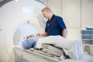
MRI
As the shoulder is a multi-layered area, an MRI is a frequently used scan for this part of the body. MRI scans can show the soft tissue as well as the bone, so this test can be especially useful in observing swelling. The MRI allows the radiologist and physician to see further detail of the interior structures. Frequently, an arthrogram is utilised to investigate shoulder pain. An arthrogram involves an injection of a contrast dye, which helps internal areas show up much more clearly on our scans. The arthrogram can be used in conjunction with a CT scan, ultrasound, or MRI.
Experiencing Pain?
There are many useful diagnostic tools and tests for determining the cause of joint pain throughout the body. Your radiologist, alongside your physician, will determine which scans are most appropriate for each patient. If you are experiencing any current joint pain, do not hesitate to contact your physician immediately. Injuries or other problems can be dealt with, and our radiology technology here at Vision XRAY is excellent for investigating these issues. Your doctor can refer you for a diagnostic scan, and together we’ll help determine the cause of your pain.
Read our articles and FAQs
We’re delighted to provide updates on the latest medical imaging technology and answer your most frequently asked questions about our services.